
AID: Bridging Clinical Microbiology and Computer Vision for Automated UTI Screening
Automatic image classification for the urinoculture screening
The paper introduces AID (Automatic Infections Detector), a fully automated system for screening Urinary Tract Infections (UTIs) using color image processing and machine learning. By utilizing multi-stage segmentation and ensembles of MLPs and SVMs, the system achieves High-performance classification across eight bacterial types and provides automated colony counting for infection severity estimation.
Executive Summary
TL;DR: The "Automatic Infections Detector" (AID) is a hardware-software integration designed to automate the diagnosis of Urinary Tract Infections. By replacing subjective human visual assessment with a robust pipeline of chromatic modeling and machine learning (MLP/SVM), AID identifies eight bacterial species and estimates infection severity through automated colony counting.
Background Positioning: This work represents a transition from traditional manual microbiology to "Digital Pathology" frameworks. It resides at the intersection of Advanced Image Processing and Clinical Decision Support Systems, moving beyond simple thresholding to handle the biological messiness of bacterial growth.
Problem & Motivation: The Bottleneck in the Petri Dish
Urinary Tract Infections (UTIs) are among the most common medical conditions globally. Despite the digital revolution, the "Gold Standard" for diagnosis remains the manual culture: seeding urine on a Petri dish, waiting 24 hours, and having a microbiologist count dots.
The authors identify three critical pain points:
- Subjectivity: Human experts vary in their estimation of bacterial load.
- Traceability: Physical dishes are hard to archive and link to digital health records effortlessly.
- Complexity: Some infections, like Candida, are nearly "invisible" to the eye because they match the color of the culture medium (UriSelect 4).
Methodology: The Multi-Stage Chromatic Pipeline
The core innovation of AID is its Multi-stage Segmentation and Hierarchical Classification strategy. Unlike a simple "one-shot" classifier, AID mimics a biologist's reasoning.
1. Robust Background Masking
Because the culture ground (agar) changes color based on nutrients and moisture, a static color filter fails. AID uses:
- CIE-Lab Space: Focusing on (a, b) components to ignore lighting fluctuations (L).
- Mean-Shift Algorithm: To cluster pixels into modal densities, helping isolate the "culture ground" from actual biological growth.
2. Hierarchical Classification
The system splits the problem into two tiers:
- Tier 1 (Macro-Pre-tagging): Distinguishes between Red (E. Coli), Blue (KES/Faecalis), and Yellow (Pseudomonas/Proteus) groups.
- Tier 2 (Intra-group Refinement): Uses specialized SVMS and MLPs to find subtle differences, such as the size of E. Faecalis vs. KES, or the "halo" presence in Proteus.
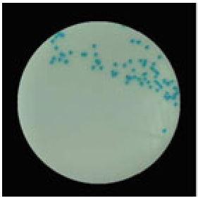 Above: Example of the GrabCut algorithm refining the isolation of colonies from the background.
Experiments & Results: Precision in the Lab
The AID system was validated on a dataset provided by DIESSE Ricerche Srl. The results highlight the power of specific feature engineering:
- Macro-Classification: Achieved 99.8% Accuracy, essentially solving the high-level identification task.
- Species-Level Accuracy: Inside the "Blue" and "Yellow" classes, accuracy hovered around 82-83%. While lower than macro-results, these figures provide a significant baseline for automated screening.
- Severity Estimation: The "Infection Severity" is calculated in UFC/ml using an ellipse-fitting algorithm that can enucleate colonies even in slightly overlapping regions.
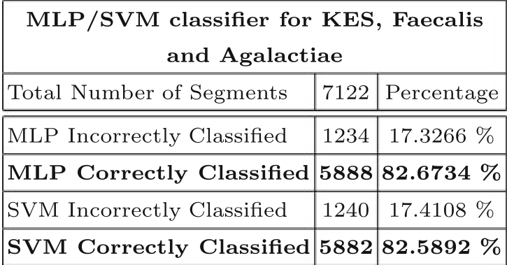 Typical performance metrics for the Blue class (KES, Faecalis, Agalactiae).
The "Invisible" Challenge: Candida
One of the most impressive aspects of the AID system is its "Ad Hoc" procedure for Candida. Since Candida is the same color as the agar, AID identifies it by looking for Extrusion Reflections. By applying a Sobel operator to find tiny edges (shadows/highlights of the 3D dome shape) and then applying a circularity filter, the system can detect colonies that are otherwise chromatically invisible.
Critical Analysis & Conclusion
Takeaway
The AID system proves that high-accuracy screening doesn't always require massive Deep Learning models; instead, a physics-informed approach to image processing (understanding how light reflects off a colony) can solve specific edge cases like Candida detection.
Limitations
- Dataset Size: The preliminary test set (35 images) is small; a larger, more diverse dataset is needed for clinical-grade reliability.
- Heavy Overlap: While the system handles "slightly overlapping" colonies, massive bacterial "carpeting" remains a challenge for automated counting.
Future Outlook
The authors plan to extend AID to transparent culture grounds and multi-enzyme substrates. As the field moves forward, integrating this with Graph Neural Networks (to model spatial relationships between colonies) could represent the next leap in accuracy.