
DeepWMA: Breaking the Robustness Barrier in White Matter Tractography Segmentation
Deep White Matter Analysis: Fast, Consistent Tractography Segmentation Across Populations and dMRI Acquisitions
The paper introduces DeepWMA, a fiber-based deep learning framework for white matter tractography segmentation. Using a novel FiberMap descriptor and a CNN classifier trained on 1 million labeled fibers, it achieves SOTA consistency in identifying 54 anatomical tracts across diverse populations and MRI acquisitions.
TL;DR
DeepWMA is a new deep learning framework that solves a long-standing problem in neuroimaging: how to consistently identify brain "wiring" (white matter tracts) across different MRI scanners and in patients with massive brain tumors. By transforming 3D fibers into a unique 2D "FiberMap," the system achieves 91% accuracy and processes a whole brain in just 8 minutes.
Context & Positioning
In the world of Diffusion MRI (dMRI), Tractography is our window into the brain's structural connectivity. However, segmenting these millions of streamlines into anatomical bundles (like the Arcuate Fasciculus) is notoriously difficult. Existing tools usually fall into two camps:
- Voxel-based (e.g., TractSeg): Fast, but gets "confused" when tumors push brain structures out of their normal place.
- Fiber-based (Clustering): High quality, but painfully slow (hours per subject).
DeepWMA sits in the "sweet spot"—it uses the robust logic of fiber-based analysis but accelerates it with Deep Learning to handle clinical edge cases that break traditional models.
The Problem: The "Messy" Brain
Why is this hard? First, a fiber can be defined from "Point A to B" or "Point B to A"—a computer needs to know it's the same thing (Order Invariance). Second, brain tumors and edema (swelling) physically displace tracts. If a model expects a tract to be at a specific coordinate, it will fail when a tumor pushes that tract 2cm to the left.
Methodology: The "FiberMap" Breakthrough
The core innovation is the FiberMap descriptor. Instead of feeding raw 1D coordinates into a network, the authors convert each fiber into a image (like an RGB image).
How FiberMap works:
- Spatial Encoding: It encodes the R-A-S (Right, Anterior, Superior) coordinates into channels.
- Invariance: By repeating and flipping coordinate sequences, the descriptor becomes insensitive to which "end" of the fiber the tracking started from.
- CNN Ready: This format allows the researchers to leverage standard, highly optimized Convolutional Neural Networks (CNNs).
 Caption: The DeepWMA pipeline: from whole-brain tractography to FiberMap generation, followed by CNN classification into 54 specific anatomical tracts.
Experiments & Results
The model was trained on 1 million labeled fiber samples from the Human Connectome Project (HCP).
1. Descriptor Comparison
The authors tested several ways to "show" fibers to the AI. Pure coordinate lists (1D-RAS) performed poorly (~47%). FiberMap hit 90.99%, proving that the 2D spatial arrangement is key for the filters to "see" the shape of the tract.

2. Clinical Robustness (The Tumor Test)
The most impressive result came from the Brain Tumor Patient (BTP) dataset. While the popular TractSeg method failed to find tracts in patients with large tumors/edema (94.4% detection), DeepWMA successfully detected >99% of tracts.
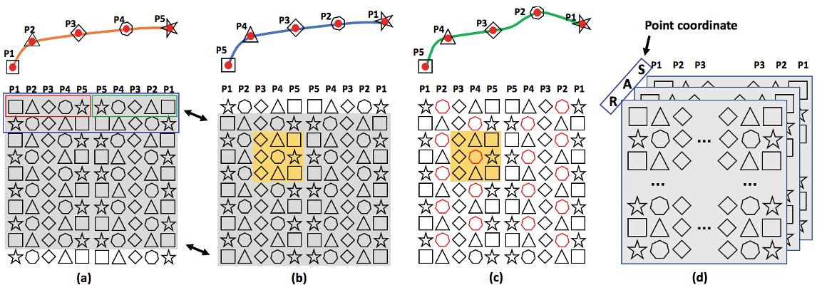 Caption: Visualizing how FiberMap handles local fiber differences due to healthy variation vs. pathological displacement.
Critical Insight & Conclusion
DeepWMA proves that representation matters. By treating a 3D streamline as a 2D image, the authors tapped into the power of Computer Vision to solve a geometry problem.
Takeaway: This is a major win for neurosurgeons. Being able to accurately map the "Corticospinal Tract" around a tumor in 8 minutes (instead of 90) means this technology can actually be used in a high-pressure clinical environment.
Limitations: The model classifies individual fibers. Future work could benefit from "contextual" classification—knowing what the neighboring fibers are doing might push that 91% accuracy even closer to 100%.
Future Outlook: We expect this "coordinate-to-map" approach to spread to other fields like vascular modeling or protein folding analysis.