
Decoding the Self: High-Accuracy Self-Esteem Recognition via Hierarchical Brain Networks
Enhancing the Representation of Multiple Anatomical Network for Young Adults with Self-Esteem Difference
This study presents a multi-level anatomical network framework designed to classify young adults based on self-esteem differences using structural MRI. By integrating four-layer hierarchical network features with ROI-based morphological measurements, the method achieves a state-of-the-art accuracy of 97.26% in distinguishing between high and low self-esteem groups.
TL;DR
Researchers have developed a sophisticated machine learning framework that analyzes the human brain as a multi-layered anatomical network to predict self-esteem levels. By moving beyond isolated brain regions and looking at how areas connect across four different scales, the model achieved a remarkable 97.26% accuracy in distinguishing individuals with high vs. low self-esteem—surpassing traditional single-network methods.
Background & Motivation: Why Single Regions Aren't Enough
Self-esteem—our fundamental evaluation of our own worth—is not "located" in a single spot in the brain. While previous research pointed toward the "Cortical Midline Structures" (like the prefrontal cortex), these studies often suffered from a reductionist view.
The pain point in current neuroimaging is the reliance on single-level network frameworks. These frameworks can't capture the subtle, "multi-scale" deviations in how the brain is structured. To truly understand self-esteem, we must look at both the fine-grained local morphology (the "nodes") and the complex, hierarchical organization of the whole brain (the "edges").
Methodology: The Four-Layer Architecture
The core innovation of this paper is the Multilevel Hierarchical Network. Instead of just looking at 78 individual regions, the authors organized the brain into four nested layers:
- Layer 1: The entire brain as a single unit.
- Layer 2: Seven major lobes (Frontal, Temporal, Limbic, etc.).
- Layer 3: Specific surfaces (Lateral, Medial, Inferior) within each lobe.
- Layer 4: 78 fine-grained cortical Regions of Interest (ROIs).
Feature Fusion & Machine Learning Pipeline
The authors didn't just dump data into a model. They used a sophisticated multi-stage pipeline:
- Feature Construction: Extracted Gray Matter volume and Cortical Thickness, alongside Pearson correlation matrices for the networks.
- Hybrid Selection: Used T-tests for initial filtering, Minimum Redundancy Maximum Relevance (mRMR) for pruning, and SVM-RFE for the final subset selection.
- Multi-Kernel SVM: A specialized classifier that uses different "kernels" to process morphological data and network data separately before fusing them based on an optimized weighting factor.
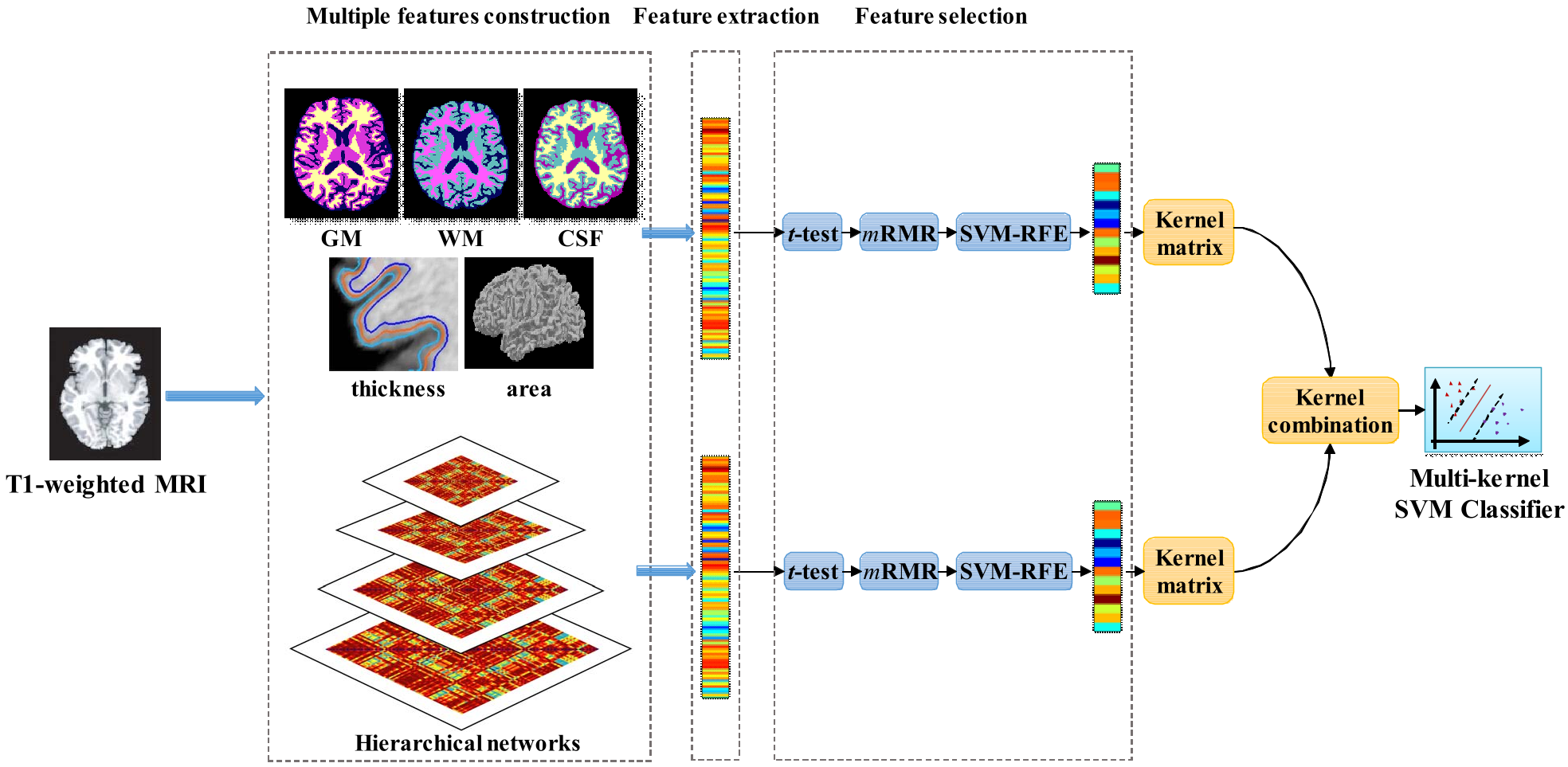 Figure 1: The proposed classification framework showing the transition from image processing to hierarchical feature extraction and multi-kernel fusion.
Experimental Results: A Significant Leap in Performance
The results provide clear evidence that hierarchy matters. As shown in the table below, the "Multilevel" approach (combining ROI features and network features across all layers) crushed the competition.
- Multilevel Accuracy: 97.26%
- Single Layer (4th Layer only) Accuracy: 90.69%
- ROI-only Accuracy: 88.69%
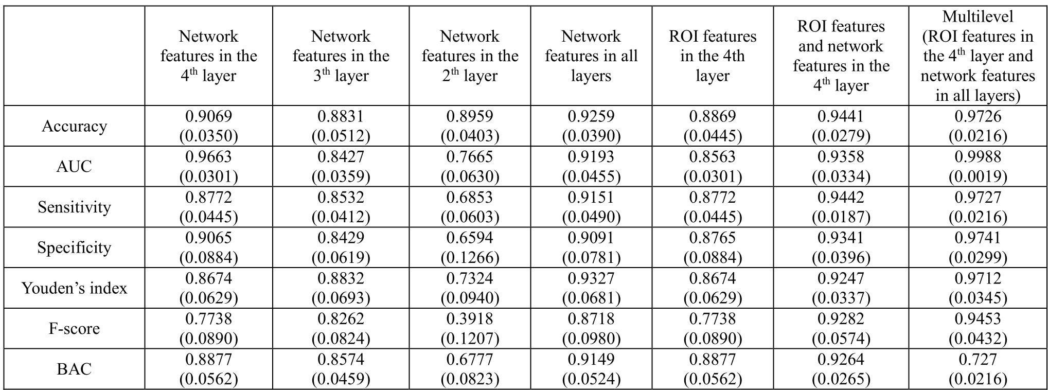 Table 2: Comparison of classification performance across different feature configurations. Note the near-perfect AUC for the multilevel approach.
Key Biological Insights
Where does self-esteem live? The model identified the Middle Frontal Gyrus, Posterior Cingulate, and Temporal Gyrus as top discriminative regions. Interestingly, it also found significant connectivity patterns between the Frontal and Limbic lobes, suggesting that the communication between our "rational" cortex and our "emotional" centers is a primary driver of self-evaluation.
Critical Insight & Future Outlook
This work demonstrates that psychological traits are "global" properties of the brain's structural architecture. The high accuracy suggests that structural MRI contains nearly all the information needed to categorize self-esteem, provided the analysis tool is sufficiently multi-dimensional.
Limitations: The study used a relatively small sample (68 subjects). While the nested cross-validation is robust, moving toward larger, more diverse datasets (e.g., UK Biobank) will be necessary to prove this model generalizes across different ages and cultures.
The Takeaway: For future AI in neuroimaging, the lesson is clear: Stop looking for the "button" in the brain and start looking at the "wiring" across scales.