
Beyond Mammals: The Veiled Chameleon’s Human-Like Embryonic Blueprint
Epiblast lumenogenesis is not a mammalian-specific trait
This study identifies that epiblast lumenogenesis, previously thought to be a mammalian-specific hallmark, occurs in the veiled chameleon (Chameleo calyptratus). By combining SEM, immunofluorescence, and in-situ hybridization, the authors demonstrate that chameleons achieve a human-embryo-like morphology through a unique "purse-string" actomyosin mechanism.
TL;DR
For decades, the hollowing of the epiblast to form a lumen was considered a unique milestone of mammalian development, separating us from the "flat" embryos of birds and reptiles. A groundbreaking study in Nature Communications (2026) shatters this view. Researchers have found that the veiled chameleon (Chameleo calyptratus) develops an epiblast lumen through an elegant "purse-string" mechanism, resulting in an embryo that looks startlingly human-like despite millions of years of evolutionary separation.
The "Flat Disc" Dogma
In developmental biology, the chicken has long been the primary model for non-mammalian amniotes. In the chick, the epiblast forms a flat, 2D disc. Consequently, science assumed that the 3D hollowing (lumenogenesis) seen in humans and mice was a mammalian "invention" necessary for implantation and amnion formation. This paper challenges that status quo by looking at a neglected corner of the evolutionary tree: the squamates (scaled reptiles).
The Mechanism: Tissue Folding and Actomyosin Cables
Unlike mammals, where the lumen forms via charge repulsion and fluid pumping, the chameleon uses mechanical force. The authors identified a purse-string-like mechanism where supracellular actin cables form concentric rings around the epiblast. These rings constrict, pulling the tissue edges together to create a vaulted 3D structure.
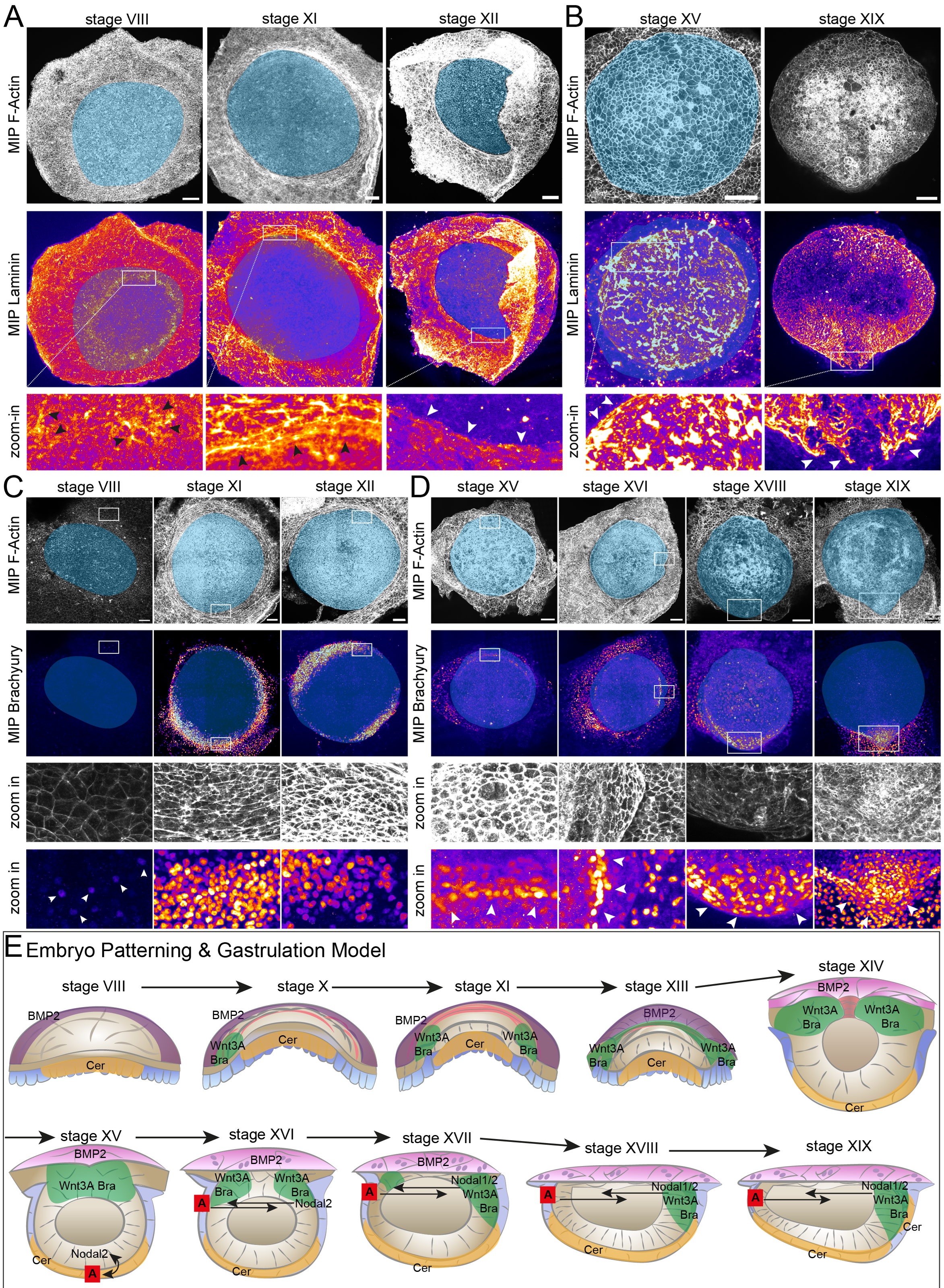 Figure: The final model illustrating the stages from flat cleavage to the closed epiblast lumen and subsequent gastrulation.
Key Molecular Findings:
- Circular Brachyury: Surprisingly, the gastrulation marker Brachyury and Wnt3a form a "dorsal ring" around the epiblast before the embryo even establishes a clear front-to-back (anterior-posterior) axis.
- Trophoblast Mimicry: The study observed binucleated cells in the chameleon's outer layer—a hallmark of the mammalian trophoblast (the part of the embryo that becomes the placenta).
Evolutionary Divergence in Patterning
While the form of the chameleon embryo mimics humans, the signaling does not. In humans and mice, genes like Cerberus and Lefty are crucial for defining the "head" of the embryo. In the chameleon, Lefty is entirely absent during these stages, and Cerberus is expressed in the "tail" region (posterior). This suggests that while nature arrived at the same 3D shape, it used a completely different set of genetic instructions to determine directionality.
Experimental Proof: Morphological Convergence
Using scanning electron microscopy (SEM) and high-resolution histology, the team compared the chameleon embryo to the human Carnegie Stage 5c. The resemblance is uncanny: both feature a dorsal trophoblast-like layer, a ventral hypoblast, and a central epiblast surrounding a hollow lumen.
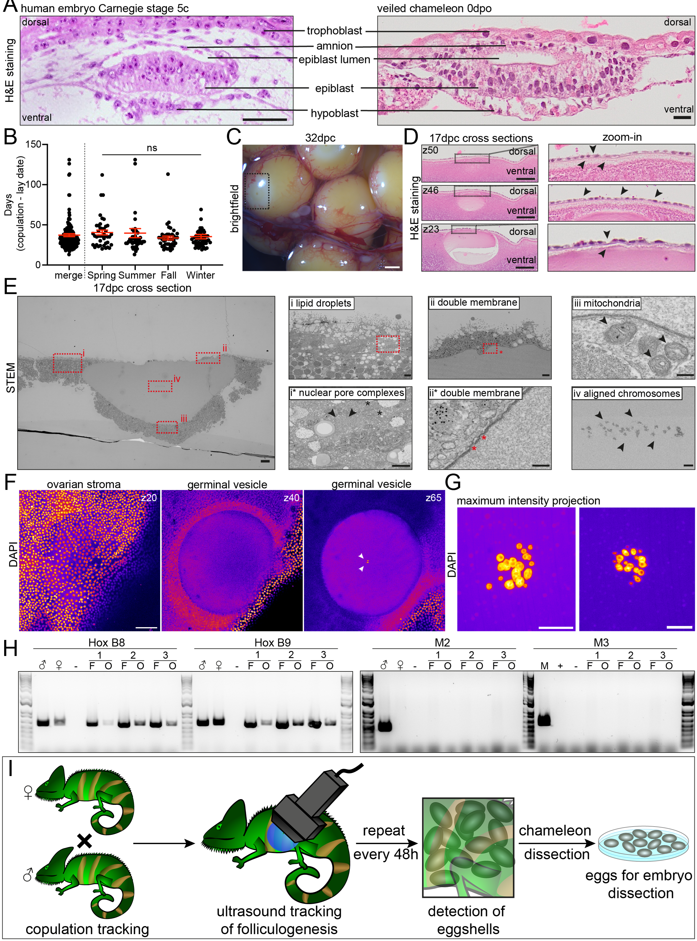 Figure: Side-by-side comparison of human (left) and chameleon (right) embryo cross-sections, showing the conserved 3D architecture.
Why This Matters
This research suggests that the "blueprint" for a 3D embryo is much older than we thought. It highlights that:
- Lumenogenesis is not mammalian-specific: It likely evolved multiple times or was lost in the lineage leading to birds.
- Mechanical Diversity: There are multiple physical ways (fluid pressure vs. actin constriction) to build a 3D embryo.
- New Models Needed: To truly understand human development, we must look beyond traditional models like the chick or mouse and explore the "evolutionary gaps" filled by species like the chameleon.
Conclusion
The veiled chameleon provides a rare window into the evolution of our own early development. By proving that 3D epiblast hollowing is a broader reptilian trait, this study forces us to rethink the origins of the amniotic cavity—the protective "aquarium" that allowed vertebrates to conquer the land.
Reference: Weberling, A., et al. (2026). Epiblast lumenogenesis is not a mammalian-specific trait. Nature Communications. https://doi.org/10.1038/s41467-026-73768-9