
Engineering the Bridge: Precise Neuronal Network Patterning on Microelectrode Arrays
Extracellular Recordings From Patterned Neuronal Networks Using Planar Microelectrode Arrays
The paper presents a methodology for reconstructing patterned hippocampal neuronal networks on planar microelectrode arrays (MEAs). By utilizing microcontact printing (μCP) and photoresist-liftoff to localize poly-L-lysine (PLL), the authors successfully recorded spontaneous bursting activity and coincident firing from precisely guided neurite bundles.
TL;DR
This research establishes a robust framework for creating "engineered" neuronal networks. By combining high-resolution chemical patterning (Microcontact Printing and Lift-off) with Planar Microelectrode Arrays (MEAs), the authors moved beyond random cell growth to create structured circuits of hippocampal neurons. They successfully recorded spontaneous electrical bursts and coincident firing from specific, guided neurite bundles, providing a roadmap for studying neural computation in vitro.
The Scalability Problem in Neurophysiology
How does network architecture shape computational function? Traditionally, neuroscientists used micropipettes (Patch-clamping), which offer incredible detail but are invasive and limited to a few cells. Conversely, early Microelectrode Arrays (MEAs) allowed for long-term, multi-site recording but lacked control over where cells grew. Without structural control, the signals on the electrodes were often an "alphabet soup" of unidentifiable sources.
The core challenge addressed here is Haptotaxis control: using surface cues to force neurons to grow into specific, predictable geometries that align perfectly with recording sensors.
Methodology: Lithography meets Biology
The authors utilized two primary techniques to deposit Poly-L-Lysine (PLL)—a "glue" that promotes cell attachment:
- Microcontact Printing (μCP): Using a PDMS "stamp" to rapidly transfer PLL patterns.
- Photoresist Lift-off: A more traditional cleanroom technique that, while time-consuming, resulted in thicker PLL layers and better long-term cell health.
To ensure the recording was viable, they addressed the Signal-to-Noise Ratio (SNR) by electrodepositing "Platinum Black" onto the gold electrodes. This process increases the effective surface area, lowering the impedance () and reducing the Johnson noise.
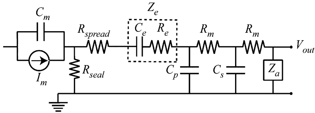 Figure 1: The circuit model (Fig 2 in paper) defines the sealing resistance () and microelectrode impedance () as the critical variables for detecting clear extracellular signals.
Key Insight: The "Duty-Cycle" of Growth
A fascinating discovery in this work is the relationship between pattern geometry and neurite "escape." The authors found that a high "duty-cycle" (where PLL lines are thick relative to the gaps) allowed neurites to wander.
- The Sweet Spot: A 4% duty-cycle (2 μm lines with 50 μm spaces) provided the best guidance, effectively "caging" the neurons into the desired architecture.
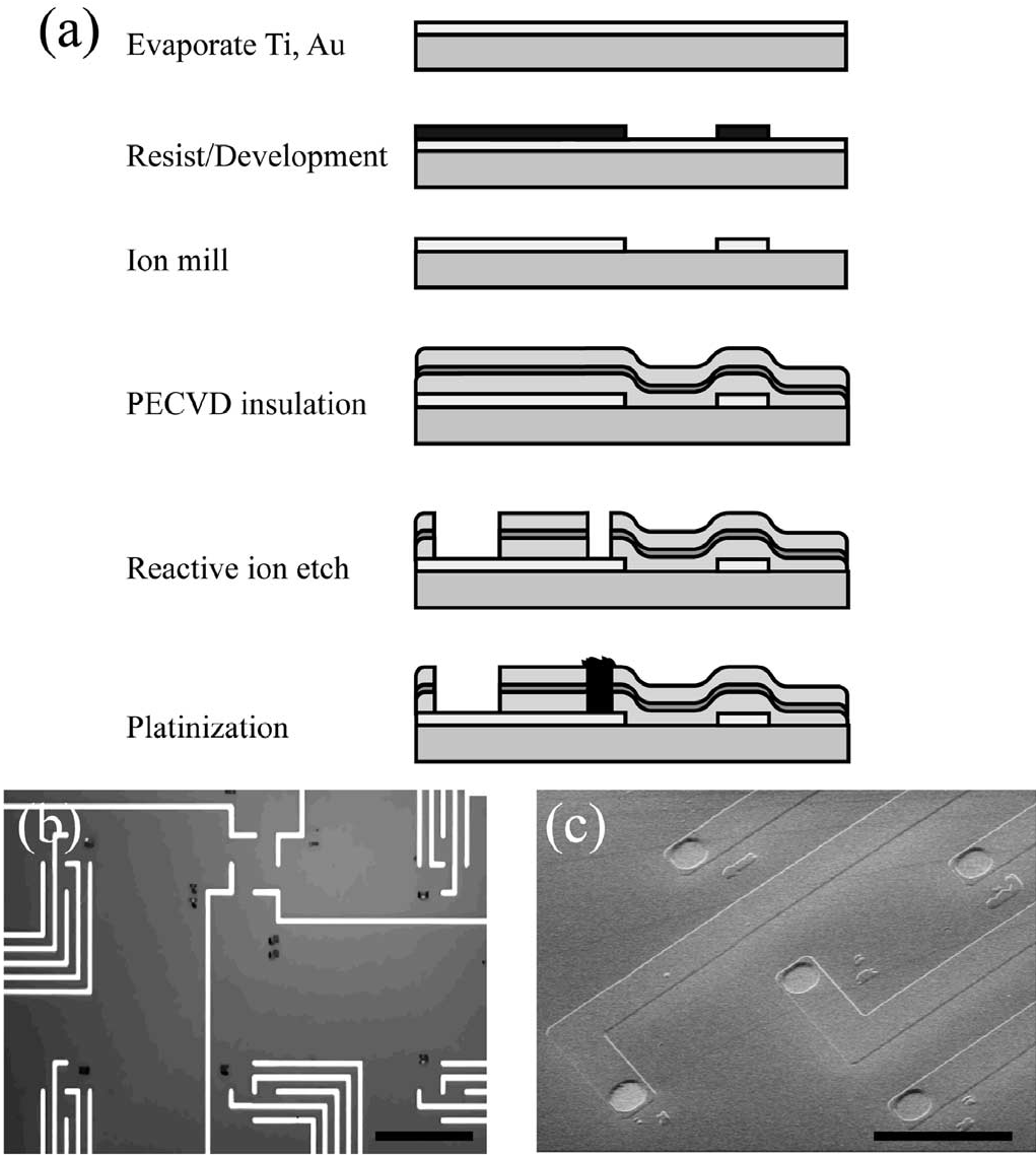 Figure 2: SEM micrographs showing the difference between a bare gold recording site and a platinized site. The increased surface area is vital for low-noise recording.
Experimental Milestones
The authors achieved several "firsts" or significant improvements in the precision of recording:
- Attenuation Observation: They recorded spontaneous bursts where the spike amplitude decreased over time—a biological phenomenon likely linked to ion channel inactivation.
- Coincident Firing: By using spike-sorting and PCA (Principal Component Analysis), they identified cases where multiple electrodes detected activity from the same cell or synchronized groups.
- Guided Bundle Recording: They successfully recorded from a 2-μm bundle of neurites, proving that the patterning wasn't just aesthetic—it was functional.
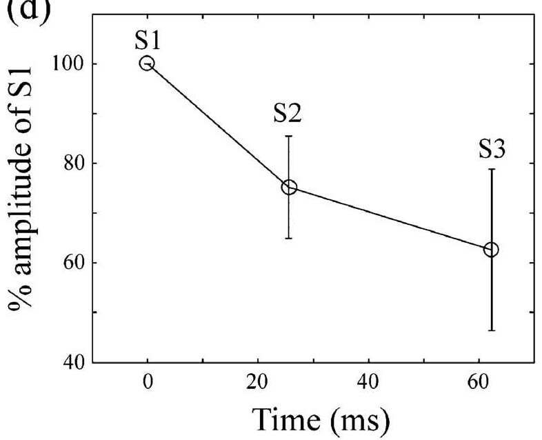 Figure 3: Phase contrast and fluorescence micrographs (Fig 5) showing the physical network and the corresponding electrical burst recordings.
Critical Analysis & Conclusion
While the paper demonstrates excellent technical mastery of MEA fabrication and patterning, it also highlights the "Biological Paradox": Neurons hate being alone.
By confining neurons to 2 μm lines (high-resolution patterning), the researchers artificially lowered the cell density. This often leads to "neurite beading" (a sign of cell stress) and lower long-term survival rates. The study concludes that moving forward, we must balance the geometrical precision required by engineers with the trophic support (cell-to-cell contact) required by biology.
This work remains a foundational step toward creating bio-hybrid computers and sophisticated "disease-on-a-chip" models where we can test drugs on structured, human-like neural circuits.