
AECRS2.0ML: Precision Cardiovascular Risk Stratification Without the Lab Work
Low-Cost Office-Based Cardiovascular Risk Stratification Using Machine Learning and Focused Carotid Ultrasound in an Asian-Indian Cohort
The study introduces AECRS2.0ML, a machine learning-based cardiovascular disease (CVD) risk calculator that integrates focused carotid ultrasound (CUS) image phenotypes with conventional clinical risk factors. Evaluated on an Asian-Indian cohort, it significantly outperforms traditional Framingham (FRS) and WHO risk scores, achieving a superior Area Under the Curve (AUC) of 0.871.
TL;DR
Researchers have developed AECRS2.0ML, an AI-driven "office-based" risk calculator that replaces expensive blood tests with focused carotid ultrasound scans. By analyzing arterial plaque morphology and clinical data through a Random Forest algorithm, the system outperformed the World Health Organization (WHO) and Framingham standards by up to 30% in an Asian-Indian cohort.
Background & Motivation: The "Resource Gap" in CVD Screening
Cardiovascular disease (CVD) remains a global leading cause of mortality, particularly in developing nations like India. Current gold-standard risk assessments (e.g., FRS) face two major hurdles:
- Economic Barriers: They require laboratory-based lipid profiles (cholesterol tests), which are often unavailable or too costly for mass screening in rural areas.
- Ethnic Blindness: Most statistical models are derived from Caucasian cohorts, making them less accurate for South-Asian populations who may have different risk profiles.
The authors hypothesized that Carotid Ultrasound (CUS)—a portable, low-cost imaging tool—could provide the "missing link" by directly measuring the biological burden of atherosclerosis (plaque) without needing a single drop of blood.
Methodology: Fusing Clinical Intuition with AI
The study utilizes a multi-step pipeline to transform raw ultrasound data into a predictive score.
1. Feature Engineering (The Integrated Approach)
Instead of relying on a single metric, the team extracted 18 features across two categories:
- Clinical Biomarkers: Age, sex, BMI, BP, and smoking status.
- Image Phenotypes: Using AtheroEdge™ software, they measured 10-year predicted values for Carotid Intima-Media Thickness (cIMT) and Plaque Area (PA).
2. The Machine Learning Engine
The core of the system is a Random Forest (RF) classifier. Unlike linear regression used in traditional scores, RF can capture non-linear interactions between risk factors—for instance, how a specific smoking history might exponentially increase risk when combined with localized plaque variability.
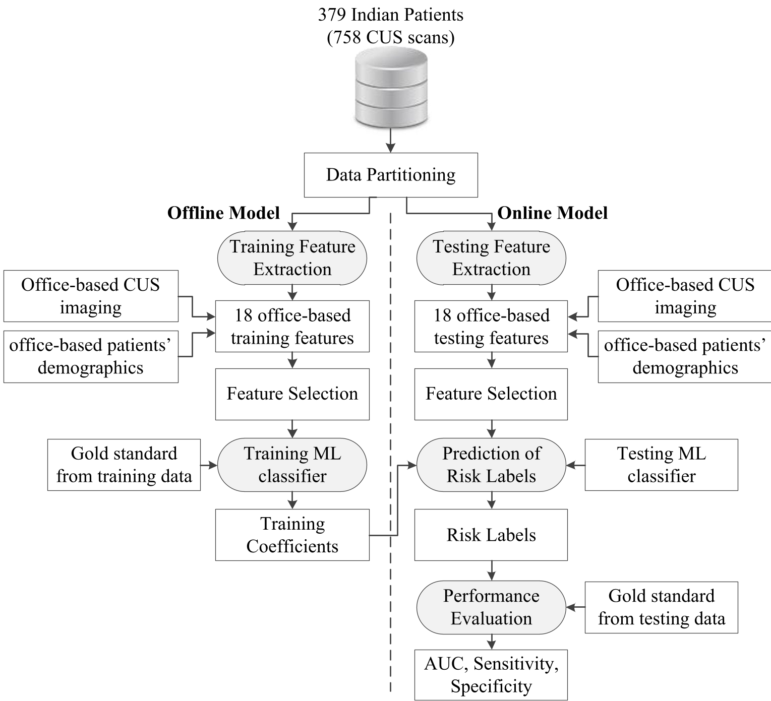 Figure 1: The AECRS2.0ML system architecture, showing the integration of ultrasound phenotypes and clinical data into the ML training pipeline.
Experiments & Results: A New Performance Standard
The model was tested on 379 patients from Chennai, India, many with comorbidities like T2DM and Hypertension.
SOTA Comparison
AECRS2.0ML decimated traditional statistics-based models:
- AECRS2.0ML: 0.871 AUC
- WHO Score: 0.727 AUC
- Framingham (FRS): 0.669 AUC
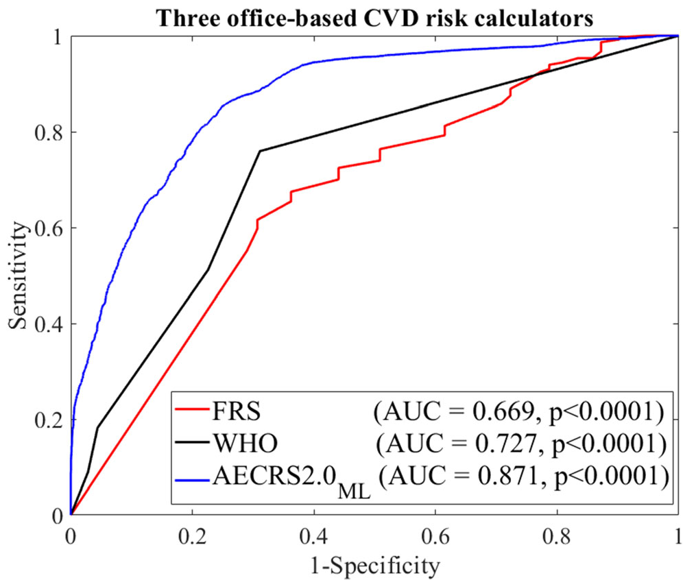 Figure 2: ROC curves demonstrating the superior discriminative power of the ML-based approach compared to FRS and WHO calculators.
The Power of PCA
The inclusion of polling-based Principal Component Analysis (PCA) improved the AUC by approximately 4%. It identified that age, systolic blood pressure, and predicted 10-year plaque area were the most critical "dominant features" for predicting risk.
Critical Analysis & Conclusion
The significance of this work lies in its practicality. By shifting from "Laboratory-based" to "Office-based" assessment, the AECRS2.0ML makes high-tier preventive cardiology accessible to underserved populations.
Limitations
- Surrogate Endpoints: The study used CKD as a surrogate for CVD rather than long-term cardiovascular event follow-ups. While pathologically linked, longitudinal data would strengthen the findings.
- Single Ethnicity: While specific to Asian-Indians (a strength for that group), the model requires further validation across multi-ethnic cohorts to ensure global stability.
Future Outlook
The next logical step is moving from Machine Learning to Deep Learning (CNNs), where the AI could automatically segment the ultrasound images, further reducing the need for expert sonographer intervention and making the tool even more "low-cost" and automated.
Takeaway: This pilot study proves that AI-empowered imaging can effectively replace expensive blood work, providing a blueprint for the future of resource-constrained healthcare.