
Decoding the Magnetic Silence: How Magnetic Fields Depress Neuronal Network Activity
Magnetic Stimulation and Depression of Mammalian Networks in Primary Neuronal Cell Cultures
The study investigates the effects of repetitive magnetic stimulation on mammalian neuronal networks using Microelectrode Arrays (MEAs). By applying biphasic rectangular pulses (0.6 T, up to 300 Hz) to primary cultures, it achieves a stable, reversible suppression of network activity, marking a significant step in quantifying the basic cellular mechanisms of Transcranial Magnetic Stimulation (TMS).
TL;DR
Researchers have successfully used primary neuronal cultures on Microelectrode Arrays (MEAs) to prove that magnetic stimulation—specifically "Magnetic Pulse Exposure" (MPE)—can significantly and reversibly suppress neuronal firing. Unlike electrical stimulation, which triggers immediate spikes, magnetic fields act slowly, likely interfering with synaptic exocytosis, offering a potential breakthrough for non-pharmacological pain management.
Context & Motivation: The TMS "Black Box"
While Transcranial Magnetic Stimulation (TMS) is widely used for treating depression and neurological disorders, it remains somewhat of a "black box." In humans, we see the macro-level behavioral changes but lack a high-resolution view of what happens at the individual synapse level within gray matter. Prior work struggled with coil heating and the sheer complexity of brain tissue.
This study pivots to in vitro neuronal networks. By growing mouse spinal cord (SC) and frontal cortex (FC) cells on MEAs, the authors created a "simplified gray matter" environment where every spike can be recorded before, during, and long after magnetic exposure.
Methodology: The Precision of Magnetic Pulse Exposure (MPE)
The team developed a custom iron-core coil to deliver biphasic pulses at flux densities up to 0.6 T. Because these cultures lack the organized geometry of large axonal tracts, the magnetic field doesn't "trigger" action potentials directly. Instead, it modulates the spontaneous rhythm of the network.
Key Experimental Setup:
- Target: Mature spontaneous networks (4 weeks+ in vitro).
- Modality: High-frequency pulse trains and burst trains (up to 300 Hz).
- Inhibition Monitoring: Used pharmacological blockers (Bicuculline) to see how the network reacts when its natural "brakes" are removed.
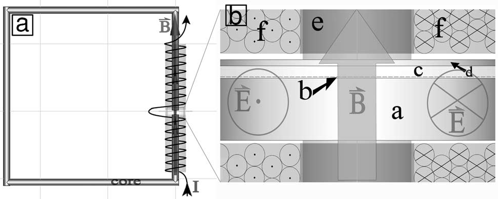 Figure: The specialized life-support and coil setup allowing for uniform magnetic exposure of the 1mm² MEA matrix.
Core Insights: Suppression over Stimulation
The most striking discovery was that magnetic pulses are predominantly inhibitory.
- Dose-Dependent Decay: As the pulse count increased from 2,500 to 15,000, the network activity decayed more significantly.
- No Immediate Response: Unlike electrical stimulation, which causes immediate firing (latency <40ms), magnetic onset took 10 seconds to several minutes. This suggests a metabolic or structural interference with synaptic release rather than a simple membrane depolarization.
- The Saturation Effect: The degree of inhibition eventually hits a plateau (saturation) determined by the frequency, not the total dose. Higher frequencies (80–100 Hz MPF) led to faster and deeper suppression.
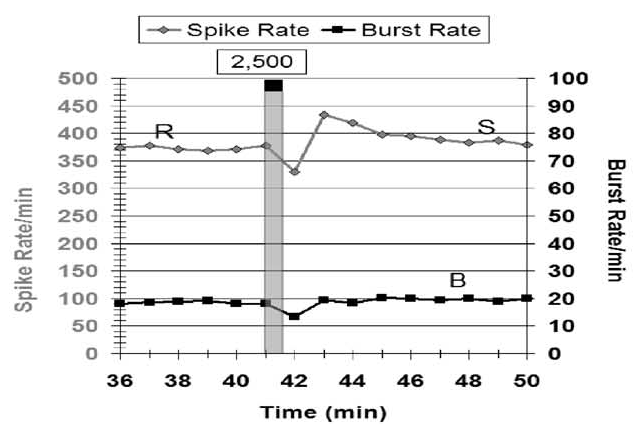 Figure: The linear relationship between pulse count and inhibitory effect, showing a gradual decay and subsequent rebound excitation.
The "Synaptic Interference" Hypothesis
Why does it happen? The authors propose that the oscillating electric fields induced by the magnetic pulses interfere with synaptic exocytosis. By observing burst patterns, they noticed that during exposure:
- Burst periods lengthened.
- Mean spike frequencies inside bursts decreased.
- The number of spikes per burst dropped.
When the researchers "disinhibited" the networks (blocking GABA receptors), they found that low-frequency pulses (10-60 Hz) could actually cause excitation. This confirms a biphasic effect: magnetic fields provide a weak excitatory push but a much stronger, overarching inhibitory pull at high frequencies.
Results & Clinical Implications
- Spinal Cord Sensitivity: SC cultures were more sensitive to suppression than FC cultures, suggesting tissue-specific responses to magnetic therapy.
- Total Suppression: At 80 Hz MPF, the team achieved 100% spike suppression with full, slow recovery.
- Pain Management: The ability to maintain 40% inhibition for over 30 minutes points toward using MPE as a non-chemical alternative to manage chronic pain.
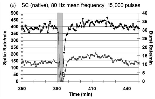 Figure: Comparison of frequency-dependent inhibition across Frontal Cortex and Spinal Cord networks under native and disinhibited conditions.
Conclusion and Future Outlook
This paper shifts the narrative from "magnetic stimulation" (as an trigger) to "magnetic modulation" (as a suppressor). By identifying the synapse as the primary target and providing quantitative data on dose-frequency limits, this research paves the way for optimized TMS protocols.
Limitations: The study is in vitro; while it mimics gray matter, it lacks the vascular and hormonal interactions of an intact brain. Future work must bridge the gap between these cellular observations and in vivo behavioral effects.