
CBMIR in Medical Social Networks: Bridging the Semantic Gap with Human-in-the-Loop AI
A medical image retrieval scheme with relevance feedback through a medical social network
The paper introduces a specialized Medical Social Network (MSN) integrated with a Content-Based Medical Image Retrieval (CBMIR) system. It leverages a fusion of low-level visual features (color, shape, texture) and a Relevance Feedback (RF) mechanism to achieve state-of-the-art precision (MAP of 0.82), specifically designed to assist physicians in diagnostic decision-making and collaborative learning.
TL;DR
This study presents a paradigm shift in medical diagnostic aids by embedding a Content-Based Medical Image Retrieval (CBMIR) system directly within a medical social network. By fusing color, texture, and shape features with an iterative Relevance Feedback mechanism, the system achieves a 20% boost in precision, helping doctors find similar past cases to validate undiagnosed examinations.
The "Semantic Gap" in Clinical Practice
In the medical world, a picture is worth a thousand words, but those words—keywords like "X-ray" or "Thorax"—are often too subjective for precise retrieval. Traditional systems suffer from the Semantic Gap: the difficulty of translating low-level visual features (like pixel intensity) into high-level medical diagnoses.
The authors argue that individual decision-making is a leading cause of medical malpractice. By creating a social network for physicians and patients, they provide a collaborative space where images can be interpreted by a "wisdom of the crowd," indexed by their actual visual content rather than just tags.
Methodology: The Power of Feature Fusion
The researchers identified that no single visual descriptor is sufficient for the diversity of medical imagery. Their solution is a robust Feature Fusion Engine:
- Color (Gray-Level): Uses global histograms and color moments (Mean, SD, Skewness, Kurtosis) to capture intensity distributions.
- Texture: Employs the Gray-Level Co-occurrence Matrix (GLCM) to calculate Haralick features like Energy, Entropy, and Contrast, capturing the granular patterns essential for identifying tissue anomalies.
- Shape: Extracts "Region Descriptors" such as Circularity, Eccentricity, and Convexity to define the boundaries of organs or lesions.
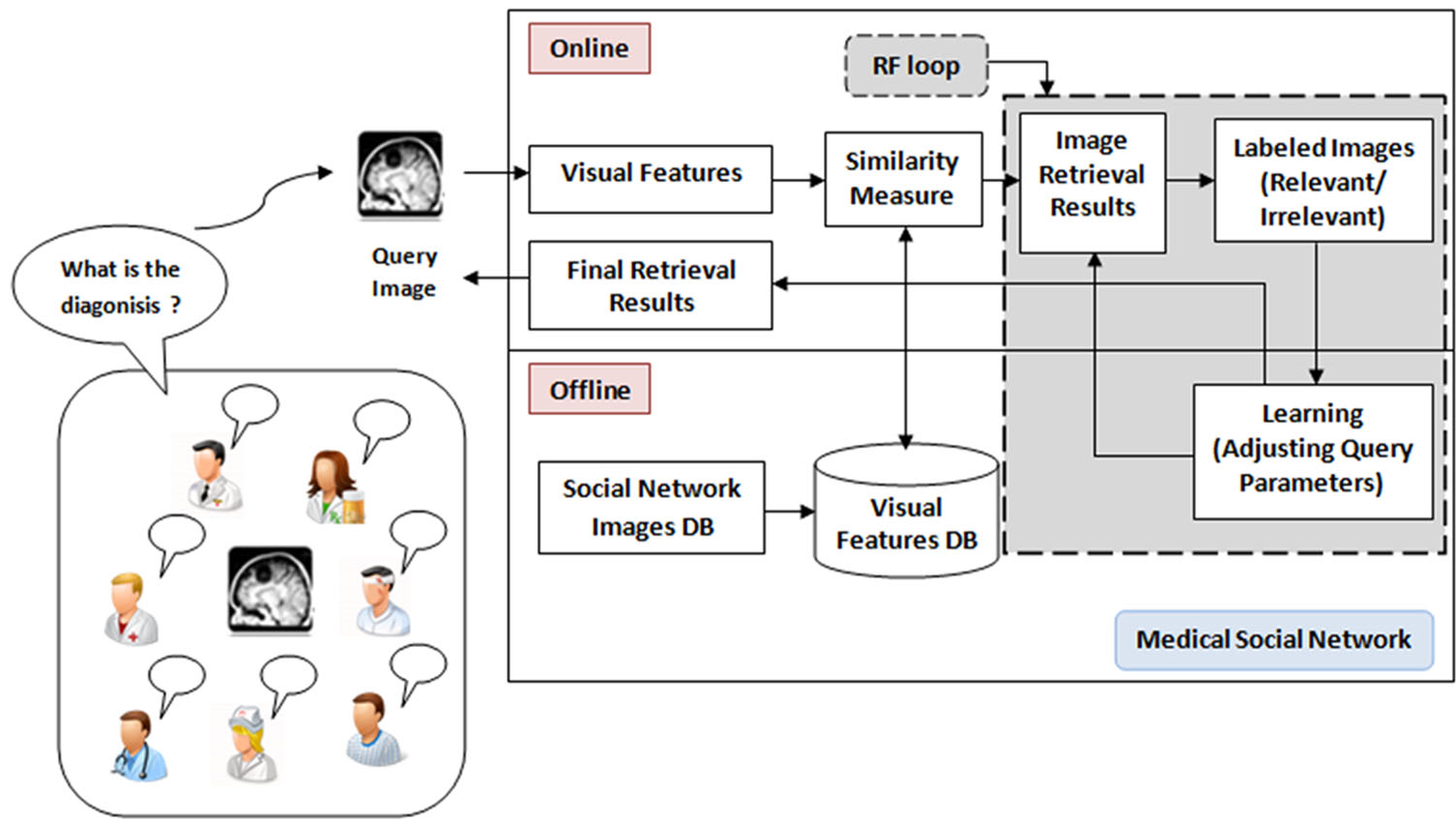
Relevance Feedback: Putting the Doctor in the Loop
The "secret sauce" of this methodology is the Relevance Feedback (RF) component. Utilizing the Rocchio Algorithm, the system allows a radiologist to mark retrieved results as "Relevant" or "Irrelevant." The system then relocates the query center in the feature space—moving toward the positive examples and away from the negative ones.
Experimental Results: Performance Breakdown
The system was tested against a dataset of 500 images across five categories: Thorax, Abdomen, Lumbar Spine, Pelvis, and Skull.
- Fusion vs. Single Feature: The results were undeniable. Using only color features for Pelvis images yielded a precision of only 30.1%. When fused with texture and shape, precision skyrocketed to 95%.
- The Power of Iteration: Each feedback cycle significantly refined the results. For Thorax images, precision improved from 69.8% to 82% over five iterations.
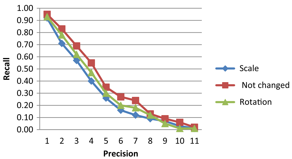 Figure: The precision-recall curves demonstrate the robustness of the system even when images are scaled or rotated.
Benchmarking against SOTA
The proposed method achieved a Mean Average Precision (MAP) of 0.82, significantly outperforming prior works by Ramamurthy (0.55/0.57) and Bueno (0.75).
Expert Acceptance & The Social Impact
Beyond the math, the authors conducted a qualitative study with 10 specialists from Charles Nicolle Hospital. The findings were highly encouraging:
- 90% of specialists believed the system helps in diagnosis and is needed in clinical routine.
- 100% found the platform easy to navigate for collaborative interpretation.
Critical Insight & Conclusion
While many researchers focus on pure "Black Box" AI, this paper highlights the value of Augmented Intelligence. By combining low-level feature fusion with the high-level expertise of physicians through a social interface, the system effectively bridges the semantic gap.
Limitations: The current system relies on "Hand-crafted" features. Future iterations would likely benefit from Deep Feature Extraction (e.g., using ResNet or Vision Transformers) to replace the manual calculation of GLCM and shape descriptors.
Final Takeaway: The integration of CBMIR into social networks transforms medical databases from static archives into "living" diagnostic tools that evolve with every physician's click.