
Bridging Radiology and Social Collaboration: A Multi-Feature CBMIR Approach
A medical image retrieval scheme through a medical social network
The paper introduces a specialized Medical Social Network (MSN) integrated with a Content-Based Medical Image Retrieval (CBMIR) system. The core method utilizes a multi-feature fusion of color (gray-level histograms/moments), texture (GLCM), and shape descriptors, achieving a Mean Average Precision (MAP) of 0.82 on clinical datasets.
TL;DR
This research tackles the high rate of medical diagnostic errors by proposing a Medical Social Network (MSN) integrated with an advanced Content-Based Medical Image Retrieval (CBMIR) system. By fusing color, texture, and shape features, the system achieves a Mean Average Precision (MAP) of 0.82, allowing doctors to find similar clinical cases and leverage past expert peer reviews for more accurate diagnoses.
Context: Beyond the Keyword Bottleneck
In modern clinical practice, the explosion of multimedia content has outpaced our ability to organize it. Traditional databases rely on Text-Based Retrieval, which is notoriously subjective—what one radiologist calls "subtle opacification," another might describe differently. More critically, the Institute of Medicine (IMNAS) estimates that medical malpractice contributes to nearly 98,000 deaths annually in the US. The solution? Moving away from isolated decision-making toward a collaborative, visual-data-driven ecosystem.
The Methodology: The Power of Feature Fusion
The authors argue that no single visual descriptor can capture the nuance of a medical image. Their solution is a robust "Feature Vector" constructed from three pillars of computer vision:
- Color (Intensity): Utilizing Gray-level histograms and moments (Mean, Standard Deviation, Skewness, Kurtosis) to capture the distribution of pixel intensities.
- Texture: Using the Gray-Level Co-occurrence Matrix (GLCM) to extract Haralick features like Energy, Entropy, and Contrast, which are vital for identifying tissue patterns.
- Shape: Employing statistical descriptors such as circularity, eccentricity, and convexity to define the boundaries of potential lesions or anatomical structures.
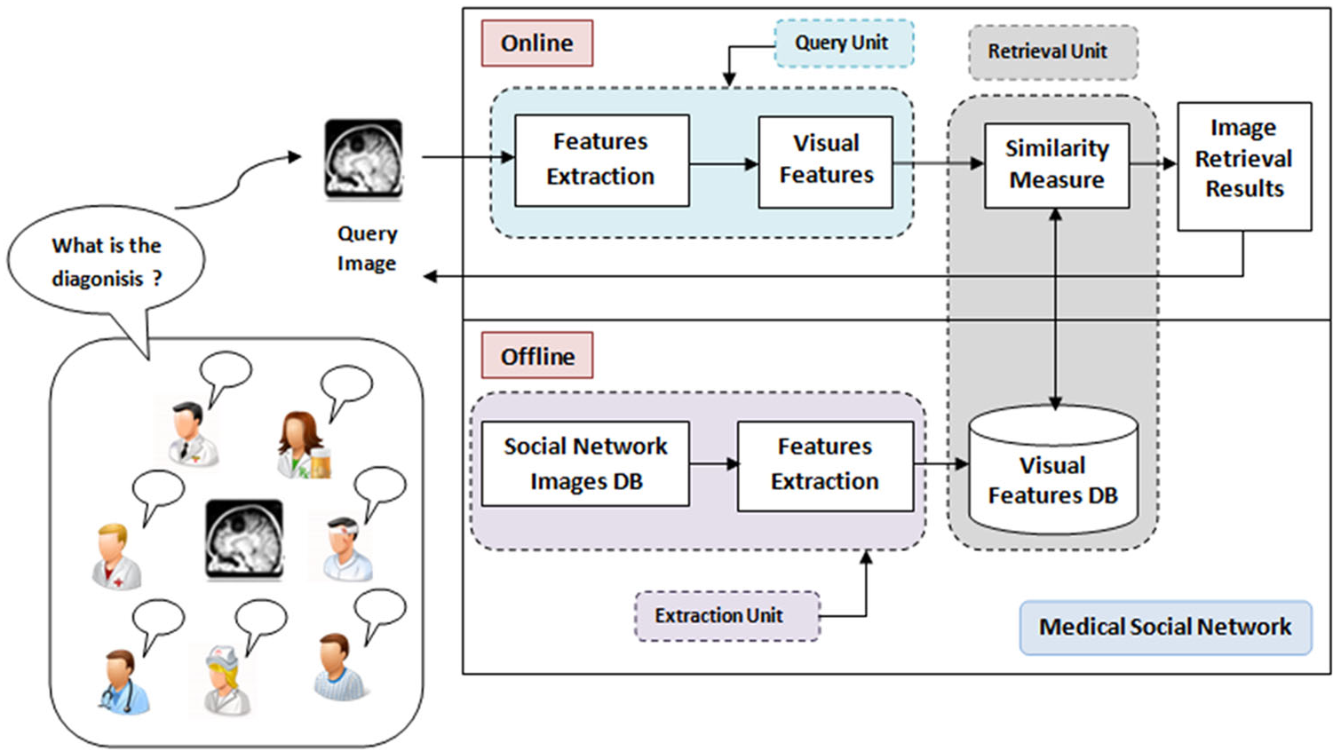 Figure 1: Conceptual structure of the CBMIR scheme within the social network.
The system employs Euclidean Distance to calculate similarity between the query image and the indexed database, returning the top 10 most visual-similar results.
Experimental Results: SOTA Performance
The system was validated using 500 medical images (Thorax, Abdomen, Skull, etc.) from Charles Nicolle Hospital. The core finding was that fusion is superior to isolation.
- The Fusion Advantage: For the "Pelvis" category, the proposed fusion method reached a precision of 0.95, while using color only yielded a meager 0.30.
- Robustness: The system maintained high performance even when images were subjected to scaling (0.9x to 1.1x) or rotation (up to 90 degrees), with only a minor 6% dip in precision.
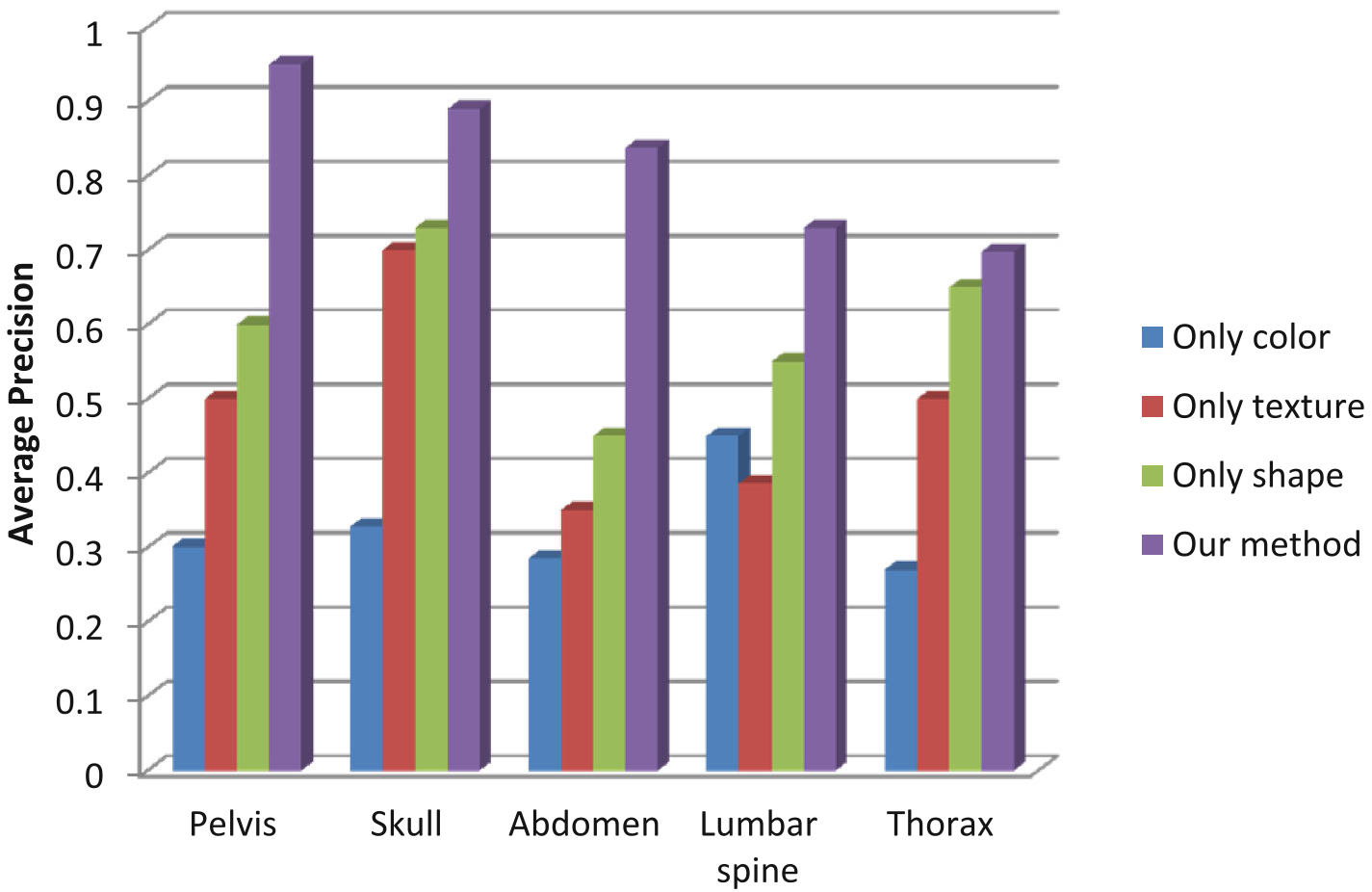 Figure 2: Average precision comparison showing the superiority of combined features over localized descriptors.
When compared to existing literature, the system's 0.82 MAP outperformed established benchmarks such as Ramamurthy and Chandran (0.57) and Bueno et al. (0.75).
Clinical Acceptance & Insights
Beyond the math, the authors conducted a survey with 10 specialists (radiologists and residents). The results were categorical:
- 90% of specialists believed the system was necessary for clinical routine.
- 90% saw it as a critical tool for training medical students.
- 100% confirmed the ease of navigation within the MSN framework.
Critical Analysis & Future Outlook
While the feature engineering is sound for its time (2016), the paper acknowledges the Semantic Gap—the disconnect between low-level visual features and high-level medical diagnoses.
Takeaway: This work underscores that the future of Digital Health isn't just about better algorithms, but about Human-AI Collaboration. By placing the "Patient at the heart of the health system" through social connectivity and empowering doctors with visual search, we can significantly reduce diagnostic uncertainty. Future iterations will likely replace manual feature extraction with Convolutional Neural Networks (CNNs) and Vision Transformers (ViT) to further bridge the semantic gap.