
The Bacterium’s Cork: How Slk Proteins Seal the Gates of Infection
Secretin-interacting plug proteins prevent antibiotic influx during type IV pilus assembly in Pseudomonas aeruginosa
2026-01-01
Summary
Problem
Method
Results
Takeaways
Abstract
The study identifies SlkA and SlkB (SlkAB) as novel "plug" proteins in Pseudomonas aeruginosa that physically seal the Type IV pilus (T4P) secretin channel (PilQ). This mechanism prevents the influx of antibiotics and maintains outer membrane integrity before the inner membrane complex docks.
## TL;DR
Pseudomonas aeruginosa faces a survival dilemma: it must open large "secretin" channels in its outer membrane to deploy virulence-associated Type IV pili, yet these same holes risk letting in lethal antibiotics. A new study published in *Nature Communications* (2026) reveals that P. aeruginosa uses two periplasmic proteins, **SlkA and SlkB**, as physical "plugs" to seal these channels until the assembly machinery is fully engaged.
## The Vulnerability of the Open Gate
Gram-negative bacteria are notorious for their robust Outer Membrane (OM), which acts as a formidable barrier against drugs. However, specialized systems like the **Type IV Pilus (T4P)** require massive pores—formed by **PilQ secretins**—to allow pili to exit the cell.
For years, it was assumed the secretin's internal gate was enough. But reality is messier: these gates are often "leaky." The researchers found that without a secondary sealing mechanism, antibiotics like erythromycin could slip through these portals, bypassing the cell's standard defenses.
## Methodology: Hunting for the "Plug"
The team used a sophisticated **Tn-seq (Transposon Sequencing)** approach, looking for mutants that became hyper-sensitive to erythromycin. They struck gold with **PA5122 and PA5123**, now renamed **SlkA and SlkB**.
### The Core Mechanism
By utilizing **Cryo-EM**, the researchers visualized the PilQ secretin with and without these Slk proteins. The difference was striking:
* **Empty PilQ**: An unobstructed conduit.
* **Slk-bound PilQ**: A dense mass occupying the central lumen, physically obstructing the path through the gate.
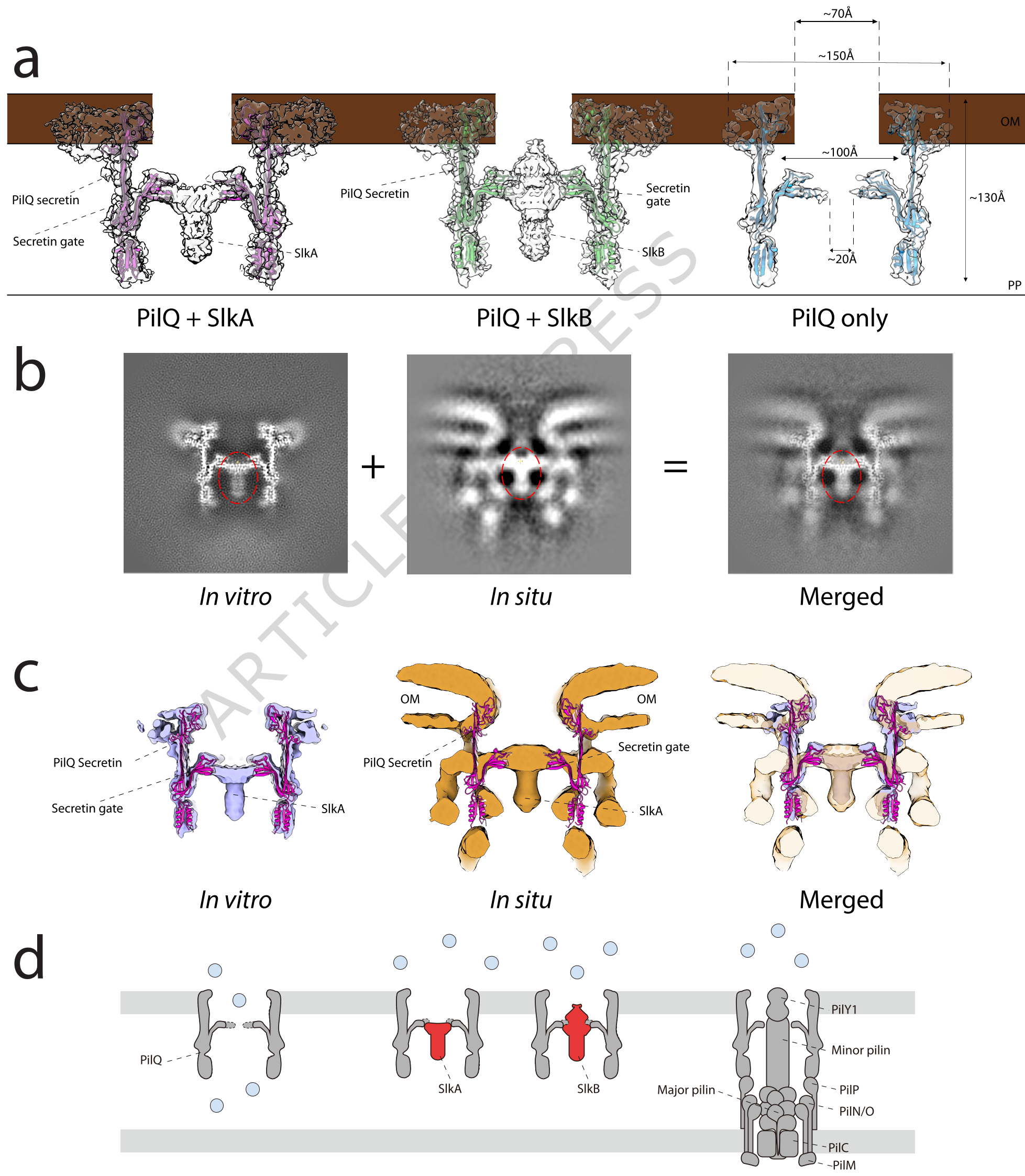
*Figure 1: Cryo-EM maps showing the Slk plugs (magenta/green) inside the PilQ channel compared to the empty state (blue).*
## Experimental Insights: Redundancy is Key
The study revealed a fascinating "Hand-off" mechanism. Slk proteins are the primary defense, but the Inner Membrane (IM) complex of the pilus assembly system also provides barrier function upon docking.
Genetic analysis showed that antibiotic sensitivity only peaked when *both* the Slk plugs and the IM complex (e.g., PilC) were disrupted. This suggests that Slk proteins serve as a "temporary cork" that stays in place until the "permanent machinery" (the pilus assembly complex) is ready to take over the space.
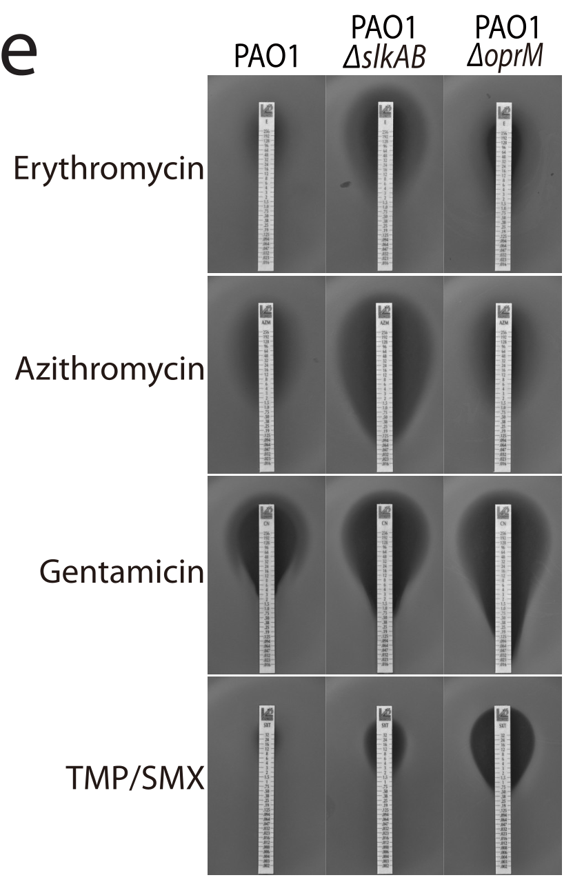
*Figure 2: Spot dilution assays showing the dramatic increase in erythromycin sensitivity when slkAB is deleted.*
## Deep Insight: A Dynamic Handoff
The most elegant part of the Slk system is its displacement. Fluorescent microscopy revealed that Slk proteins localize to the poles (where PilQ resides). Crucially, this localization **increases** when the IM complex is missing.
**The Physical Intuition**: Slk proteins and the IM complex compete for the same spot. When the pilus starts growing, it pushes the Slk "plug" out. This ensures the barrier is never left unattended—a "fail-safe" for bacterial integrity.
## Conclusion & Future Outlook
SlkAB represents a previously hidden layer of bacterial defense. By understanding how these plugs work, we open a new front in antibiotic development. If we can develop a drug that prevents SlkA/B from binding to the secretin, we could effectively "leave the door open," allowing traditional antibiotics to flood in and kill otherwise resistant P. aeruginosa.
### Takeaway
The OM barrier is not just a static wall; it is a managed interface where specialized proteins like SlkAB act as gatekeepers, ensuring that the machinery of infection doesn't become the gateway to the bacterium's own destruction.