
Beyond the Isolated Voxel: A Field Theory Revolution in Medical Imaging
14928_A Tracer-Kinetic Field Theory for Medical Imaging.
This paper introduces a Tracer-Kinetic Field Theory, a novel mathematical framework for analyzing dynamic medical imaging data (DCE-MRI, PET, CT). It shifts from the traditional isolated-voxel approach to a continuous field model, achieving superior accuracy in measuring perfusion and permeability by accounting for spatial interactions between neighboring voxels.
TL;DR
Tracer-kinetic modeling is the "math engine" behind functional MRI and CT. For decades, we have treated each pixel as an island. This paper by Steven Sourbron replaces that 1D temporal view with a 3D Field Theory. By modeling how blood and tracers flow between pixels using convection and diffusion physics, it eliminates the need for external Arterial Input Functions (AIF) and corrects long-standing errors in perfusion measurement.
The Problem: The "Isolated Island" Fallacy
In typical Dynamic Contrast-Enhanced (DCE) imaging, we measure how a contrast agent enters and leaves a tissue. Current SOTA methods assume:
- Every voxel is an isolated system.
- Every voxel is fed by one Global Arterial Input Function (AIF).
Why is this wrong? First, the AIF is usually measured in a large artery far from the tissue, leading to bolus dispersion (the pulse of contrast "smears" as it travels). Second, a single voxel often contains a mix of large feeding vessels and tiny capillaries. Traditional models can't distinguish between blood just "passing through" a large vessel and blood actually "feeding" the tissue (perfusion).
The Insight: From ROIs to Fields
The author proposes that instead of looking at time curves in isolation, we should view indicator concentration as a unified field.
Core Methodology: The PDE Framework
The theory is built on the local law of indicator mass conservation:
abla} \cdot \vec{j}$$ Where $\vec{j}$ represents the flux density. The paper elegantly breaks this down into three physical transport mechanisms: 1. **Convection**: Transport by the velocity of blood flow ($\vec{u}$). 2. **Diffusion**: Molecular movement driven by concentration gradients ($D$). 3. **Exchange/Decay**: Moving between compartments (e.g., from plasma to interstitium). 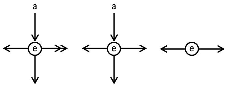 *Fig 1: Field models for weakly vascularized tissues, moving from complex molecular diffusion (left) to simplified pseudo-diffusion (right).* ## Key Achievement: Redefining Perfusion One of the most striking contributions is the mathematical definition of perfusion ($F$). In the field theory, perfusion is the **negative divergence of the arterial flow field**: $$\boldsymbol{F} = - \vec{ abla} \cdot \vec{f}^a$$ This is a "Eureka" moment for medical physics: it proves that "bypass flow" through an ROI (where in-flow equals out-flow) results in zero perfusion because the divergence is zero. This naturally filters out macrovessel contamination. ### Multi-Compartment Interaction The paper extends this to complex tissues by coupling arterial ($a$), venous ($v$), and extravascular ($e$) spaces. 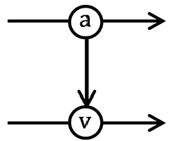 *Fig 2: A three-compartment field model accounting for blood flow, perfusion, and capillary permeability.* ## Experiments and Implications While primarily a theoretical paper, it provides a roadmap for a "Boundary Value Problem" approach to imaging: - **No more AIF**: Since voxels are coupled, the concentrations at the *boundaries* of the image scan provide the only necessary inputs. The system becomes so overdetermined that you can solve for the physics of the blood flow without needing a separate arterial measurement. - **New Biomarkers**: We can now measure the **directionality** of blood flow and interstitial fluid pressure—critical factors in how drugs reach the center of a tumor. ## Critical Analysis & Future Work **Limitations**: The primary challenge is computational. Solving a sparse, non-linear global optimization problem for millions of voxels simultaneously is significantly harder than fitting a single-compartment model. **Takeaway**: This paper moves medical imaging closer to **Computational Fluid Dynamics (CFD)**. It treats the human body's microcirculation as a coherent physical system rather than a collection of independent pixels. As GPU computing power increases, "Field Theory Analysis" will likely become the standard for high-precision cancer imaging and neuro-profiling. --- **Author Note**: This work marks a shift from "Data Fitting" to "Physics-Informed Reconstruction" in clinical radiology.