
Deciphering Atrial Mechanics: A 3D Finite-Element Approach to Wall-Motion Heterogeneity
Wall-Motion Based Analysis of Global and Regional Left Atrial Mechanics
This paper introduces a novel 3D biomechanical modeling framework for assessing Left Atrial (LA) mechanics by combining Cardiac Magnetic Resonance (CMR) imaging with finite-element surface fitting. The method utilizes a Newton optimization scheme to reconstruct continuous endocardial surfaces, enabling the quantification of both global volume changes and regional wall motion heterogeneity in healthy individuals and Atrial Fibrillation (AF) patients.
TL;DR
Researchers have developed a precision-engineering approach to mapping the heart's left atrium. By fitting a continuous finite-element "skin" to CMR data, they can now quantify exactly how different regions of the atrium contract and relax. This method reveals that Atrial Fibrillation (AF) doesn't just weaken the heart—it makes its motion physically disorganized and "stuttered" across different surface regions.
Background: The Resolution Dilemma
The left atrium is a difficult target for biomechanical analysis. Its walls are paper-thin (often just 2mm), making standard tracking techniques like CMR tagging nearly useless. Traditionally, clinicians look at "Ejection Fraction"—a single number representing global volume change. However, AF is a disease of heterogeneity. It creates patches of scar tissue and electrical "stretching" that vary wildly from the roof of the atrium to the mitral valve. To understand AF, we need a map, not just a number.
The Engineering Insight: Beyond 2D Slices
The authors moved away from simple 2D measurements, instead treating the atrium as a dynamic 3D manifold. Their core innovation lies in the Finite-Element Surface Fitting.
1. The Dynamic Coordinate System
One of the biggest hurdles in cardiac modeling is that the heart moves "vilely" within the chest—it rotates, translates, and tilts. The team established a coordinate system anchored by the Pulmonary Veins (PV) and the Mitral Valve (MV).
- The PV plane remains relatively stable (the anchor).
- The MV plane acts like a piston (the driver). By using a "floating origin" that moves with this piston, the researchers could filter out the "noise" of the heart moving through space and focus purely on the "signal" of the atrial wall shortening.
2. Mathematics of the Surface
The team utilized Cubic Hermite elements. Unlike simpler linear models, these ensure continuity—meaning the surface and its slopes are smooth at every boundary. This is critical for calculating regional strain accurately.
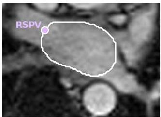 Figure 1: From raw CMR contours (g) to a reconstructed finite-element mesh (h) that accounts for tilting and translation.
Experimental Results: Mapping the Dysfunction
The model was tested on 23 subjects (healthy volunteers vs. AF patients).
- Fitting Precision: The system achieved an RMSE of 2.3mm. In the world of clinical imaging, where pixel sizes are often 1.2mm, this represents an incredibly tight fit to the actual anatomy.
- Discovery of Regional "Stutter": While healthy hearts showed a synchronized "passive-then-active" emptying phase, AF patients showed temporal desynchrony. Using 2D "Hammer Projections" (maps that flatten the sphere into a rectangle), the researchers visualized how AF patients have significant variation in when different parts of the atrium contract.
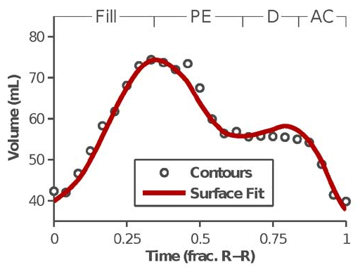 Figure 2: Hammer Projections mapping active contraction. Red/Yellow zones indicate higher fractional shortening, while Blue zones indicate mechanical stagnation.
Critical Insights: Why This Matters
The clinical value here is Ablation Targeting. Currently, surgeons scar (ablate) the atrium to stop AF, but they often do so blindly regarding mechanical impact.
- Selective Preservation: If a specific region (like the inferior wall) is shown to provide 70% of the mechanical "kick," surgeons might choose a different path to avoid scarring that specific zone.
- Disease Staging: Mechanical heterogeneity could serve as a more sensitive biomarker for disease progression than simple volume expansion.
Limitations & The Path Ahead
The primary bottleneck is manual labor. It currently takes 6–8 hours to manually contour the images for one patient. For this to reach the ER or the surgical suite, we need AI-driven auto-segmentation to feed the finite-element solver. Additionally, the study required patients to be in sinus rhythm during the scan; future work must address the "chaotic" motion of a heart currently in active fibrillation.
Conclusion
By treating the heart as a geometric problem solvable via Newton optimization, Moyer et al. have bridged the gap between raw clinical images and actionable biomechanical data. The ability to "see" mechanical desynchrony in 3D opens a new frontier for personalized cardiovascular therapy.