
Decoding the Aging Brain: Beyond Simple Diffusion Tensors
White matter microstructure across the adult lifespan: A mixed longitudinal and cross-sectional study using advanced diffusion models and brain-age prediction
This study presents a comprehensive analysis of brain white matter (WM) microstructure across the adult lifespan (18–94 years) using a mixed longitudinal and cross-sectional design. By comparing advanced multi-shell diffusion MRI (dMRI) models—including DKI, NODDI, RSI, SMT-mc, and WMTI—against conventional DTI, the authors achieve SOTA age prediction accuracy (r=0.85) and identify specific microstructural metrics with superior age sensitivity.
TL;DR
How does our brain's "wiring"—the white matter—actually change as we age from 18 to 94? This study moves beyond the classic, somewhat "blunt" Diffusion Tensor Imaging (DTI) and utilizes advanced multi-shell diffusion MRI models (like NODDI and RSI) to map the microscopic landscape of the adult lifespan. By combining these models with machine learning, researchers reached a high level of accuracy in predicting biological "brain age" and identified specific metrics that are far more sensitive to aging than the traditional FA (Fractional Anisotropy).
The Problem: The "Black Box" of DTI
For decades, DTI has been the workhorse of neuroimaging. It tells us that water diffusion becomes less "directional" as we age. But why? Is it because axons are disappearing? Is the myelin sheath thinning? Or are the fibers simply becoming more tangled (orientation dispersion)?
Traditional DTI cannot answer these "Why" questions because it treats the tissue in a voxel as a single, simplified compartment. To fix this, the authors turned to Tissue Models, which mathematically split the MRI signal into different biological pools—like the water trapped inside axons versus the water in the surrounding space.
Methodology: A Multi-Model Showdown
The researchers didn't just pick one method; they compared six:
- DTI & DKI: Signal-based models (the "Old Guard").
- NODDI & WMTI: Tissue-based models focusing on neurite density and axonal integrity.
- RSI & SMT-mc: Advanced techniques designed to handle complex fiber crossings and "slow" diffusion compartments.
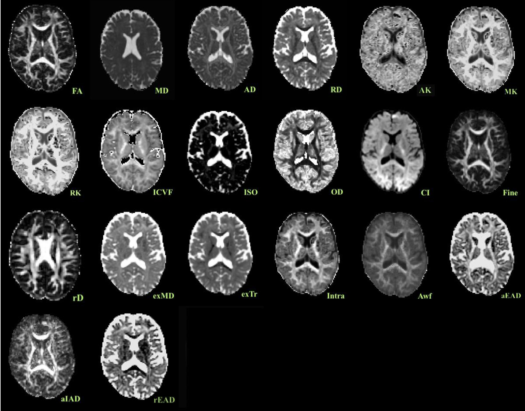 Fig 1: A visual comparison of the different metrics extracted from a single participant. Note the high detail in advanced metrics like RSI and NODDI compared to standard MD.
Using Linear Mixed Effects (LME) models, they analyzed both cross-sectional data (comparing different people) and longitudinal data (tracking individuals over ~15 months).
Key Insights: When Does the Decline Start?
The study found that "age labels" are not linear. Most white matter metrics follow a curvilinear path.
- The Turning Point: For many metrics, the peak of "brain health" occurs in the 30s or 40s.
- The Winners: The "FA fine" scale from the RSI model and the "Orientation Dispersion (OD)" from NODDI were the most sensitive markers of aging.
- The Machine Learning Edge: When all 420 features from these models were fed into an XGBoost regressor, the "Brain Age Gap" became much clearer, showing a correlation of 0.85 with actual age.
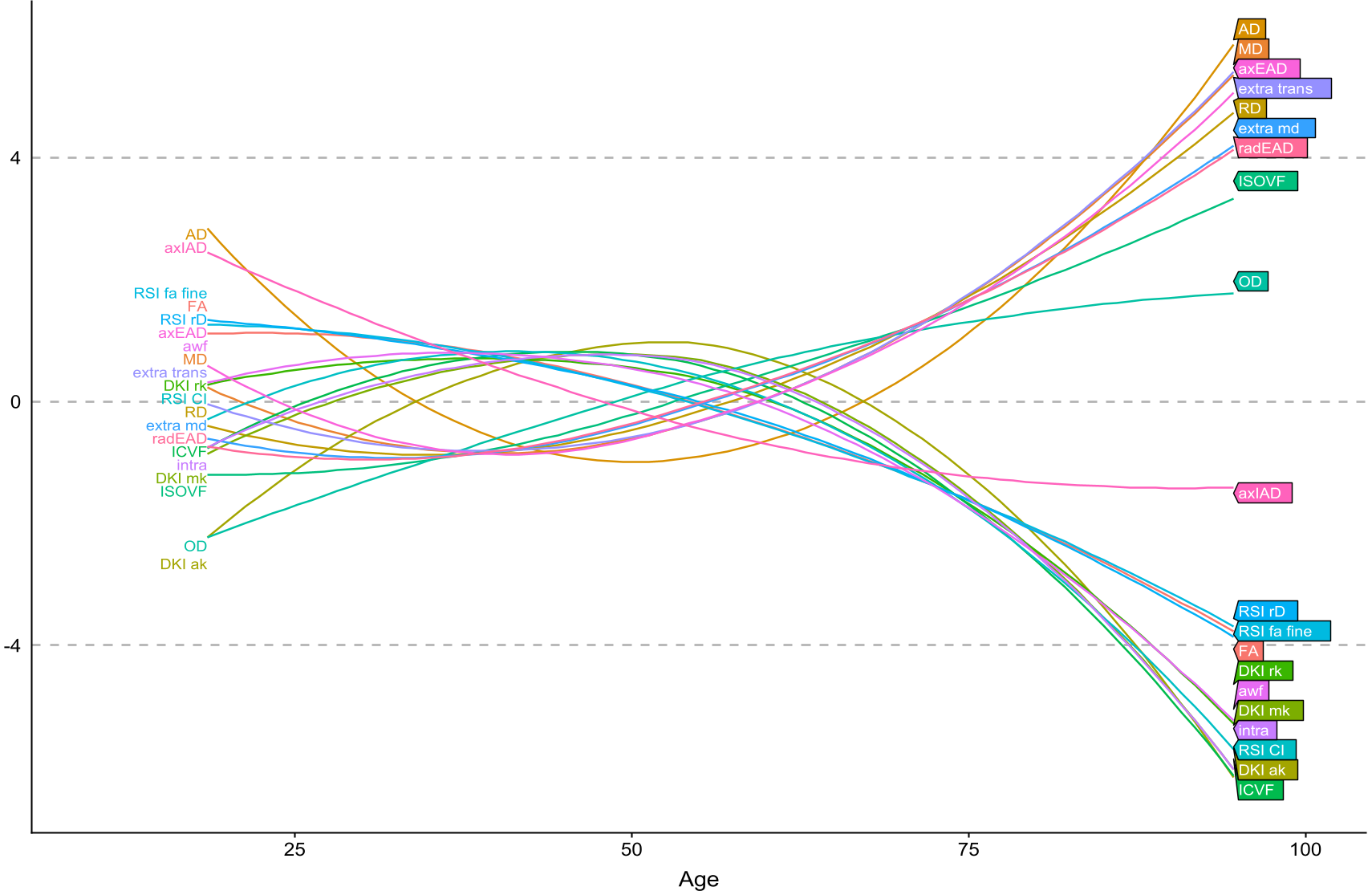 Fig 2: Standardized age curves for multiple metrics. Notice how metrics like 'OD' (Orientation Dispersion) rise steadily, while 'AWF' (Axonal Water Fraction) peaks and then drops, highlighting different biological processes.
Why This Matters
This isn't just about knowing how old your brain looks. By identifying which specific microstructural components change (e.g., increased dispersion vs. decreased axonal density), we can:
- Develop better biomarkers for early-stage dementia or multiple sclerosis.
- Understand "Super-Agers": Why do some 80-year-olds have the white matter integrity of a 50-year-old?
- Refine Clinical Trials: Use these sensitive metrics to see if a new drug is actually protecting the "wiring" of the brain.
Critical Analysis & Future Work
While the study is robust, the authors admit that longitudinal intervals were relatively short (15.2 months). Aging is a slow process, and longer follow-ups are needed to truly separate cohort effects from individual aging. Furthermore, while these models are "biophysically inspired," they still rely on mathematical assumptions that need further validation with histology (microscope-level study of real tissue).
In the future, the integration of these "advanced fingerprints" into routine clinical scans could transform how we monitor brain health throughout life.
Conclusion
The move from "signal models" to "tissue models" marks a paradigm shift in neuroimaging. This study proves that the extra complexity of multi-shell MRI pays off, providing a higher-resolution view of the aging human connectome.